Technology
We are pleased to provide a brief description of the advancements offered by our dental office.
ELECTRIC HANDPIECES
Due to increased efficiency through design, most procedures are completed in a fraction of the time as opposed to using air driven handpieces. This provides for shortened appointments and a more efficient use of time.
CEREC (Chairside Economical Restoration of Esthetic Ceramics)
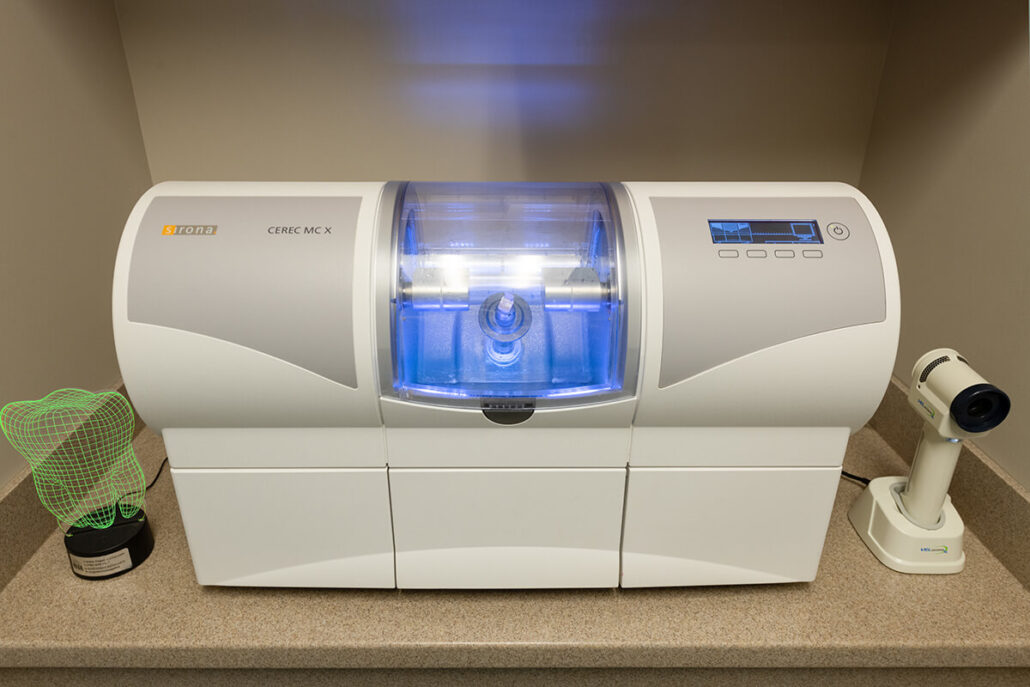
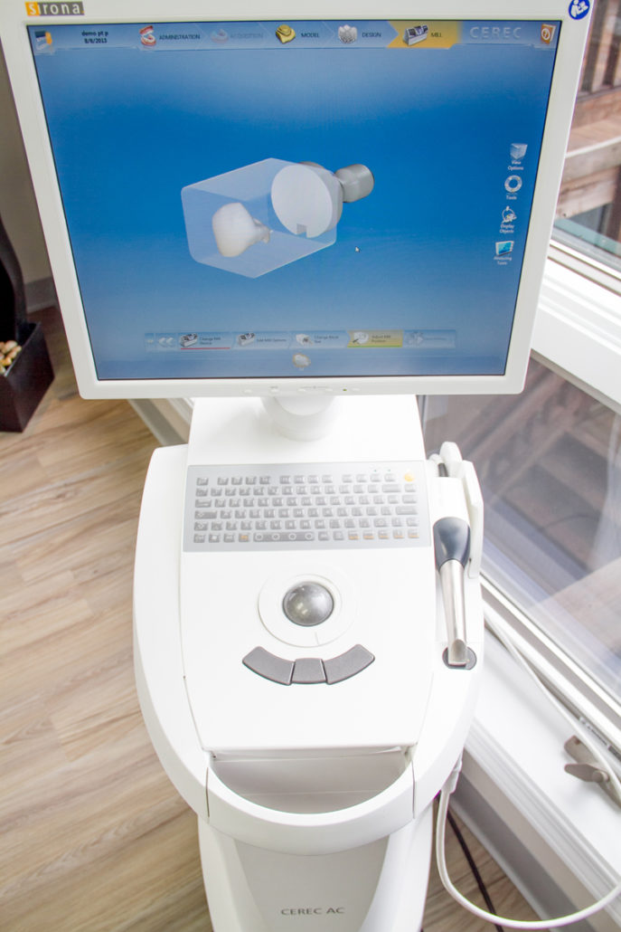
This enhanced chairside system allows for the completion of crowns in a single visit, without the use of impression trays or provisional (temporary) crowns, and only one series of injection of local anesthesia. Typically, the prepared tooth is scanned using the CEREC Omnicam, and a three dimension digital model is created. Proprietary software is then used to create a restoration using comparisons of the surrounding teeth. The dentist then refines the model using 3D CAD software. When complete, a milling chamber carves the actual restoration out of a ceramic block (lithium disilicate), using diamond head burs under computer control. The restoration is then adjusted intraorally, and subsequently placed in a sintering furnace to achieve maximum strength. The final restoration is biocompatible and highly aesthetic.
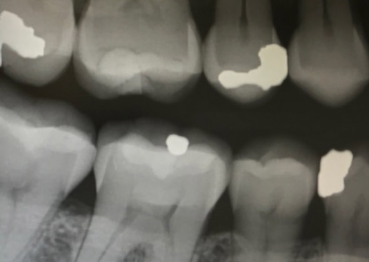
DIGITAL RADIOGRAPHY
The dental technician places a small sensor inside the patient’s mouth and an image is acquired, instantly appearing on a computer screen in the exam room. The digital image may be enlarged and manipulated, allowing dental personnel to better evaluate and detect pathology. The patient receives substantially less radiation than traditional x-rays, and we are helping our environment by eliminating the use of potentially hazardous chemicals used in the developing of traditional film.
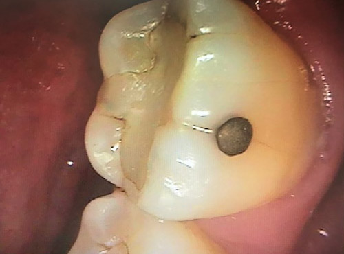
INTRAORAL CAMERA
A camera is placed intraorally and acquires images of the oral structures, which are then projected on a computer screen. By visualizing the images on a chairside monitor, patients gain a better understanding of existing conditions and the staff is able to accurately make the proper diagnosis and treatment plan. These images are stored in our computer system, and may be used by specialists or for insurance purposes.
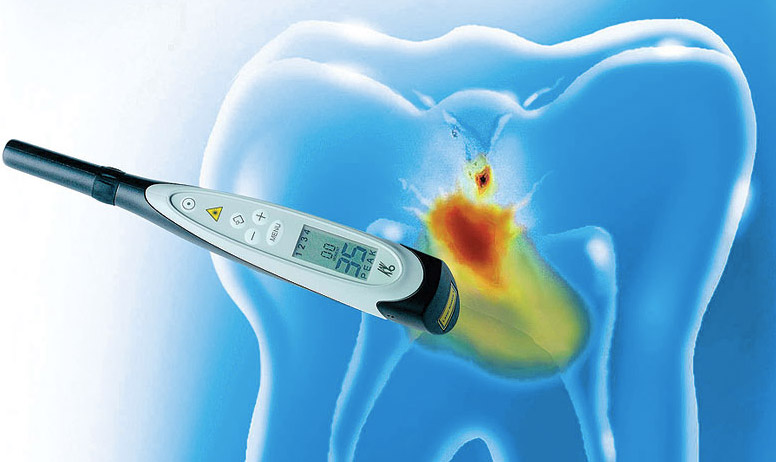
DIAGNODENT
The Diagnodent is a laser fluorescence cavity detection aid which measures changes in the tooth structure caused by caries. It is over 90 per cent accurate in detecting early cavities, especially in the pit and fissure areas on the biting surface of teeth. It is noninvasive and pain free. By using a number scale and audible alert, it can detect even the smallest carious lesion, and thus allows Dr. Rogol to preserve as much healthy tooth structure as possible by conservative treatment. The Diagnodent has received the Seal of Acceptance by the American Dental Association.
VELSCOPE
More than 480,000 new cases of oral and throat cancers are diagnosed each year, and over 35,000 of those cases are in the United States. Oral cancer kills one person every hour in the US, and without proper detection methods, a person may have oral cancer, and not even be aware of it. Early detection and treatment play a major role in the patient’s chance of survival.
Risk factors include for oral cancer include;
- frequent use of tobacco products
- excessive consumption of alcohol
- genetics (a family history of cancer)
- compromised immunity system
- excessive exposure to sun at a young age
- presence of HPV (human papillomavirus)
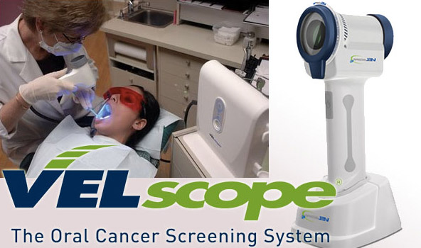
The Velscope system emits a light into the oral cavity which causes the tissues to fluoresce. Abnormalities appear much darker under the light, allowing the practitioner to detect early changes in the cellular structure. The examination is painless, noninvasive, and does not involve stains or rinses. The procedure is approved by the Federal Drug Administration (FDA).
